High risk groups 1,2
- Those living in rural and remote communities
- Aboriginal and Torres Strait Islander adults and children
- Over 75 years of age
- Those with established lung diseases
- Those with cystic fibrosis, Kartagener’s syndrome and primary ciliary dyskinesia
- Those with Chronic obstructive pulmonary disease
- Rheumatoid arthritis
Urgent referral
- For an acute exacerbation refer to the Primary Clinical Care Manual
Special considerations
- Those with cystic fibrosis are managed by a specialist
1. What is bronchiectasis? 1–5
- A chronic lung condition, defined as the permanent dilatation of the bronchi and bronchioles where the elastic and muscular tissue is destroyed by re-occurring inflammation and infection
- The damage impairs the natural drainage of bronchial secretions resulting in airway obstruction and progressive lung damage characterised by persistent:
- cough
- sputum production
- recurrent respiratory infections
- Symptoms may occur for many years before a diagnosis is confirmed
- Haemophilis influenzae and Pseudomonas aeruginosa pathogens are a primary cause of bronchiectasis airway infections
- Nearly 2% of Aboriginal and Torres Strait Islander children will develop bronchiectasis
- No definite cause can be established in up to half of all patients
- Up to 50% of patients will also have Chronic obstructive pulmonary disease
2. Diagnosis of bronchiectasis 1–5
- Bronchiectasis relies on both a clinical and radiological diagnosis
- Predicting mortality and exacerbation rates in bronchiectasis can be undertaken with an online bronchiectasis prediction tool. See Resource 1.
Flowchart 1. Diagnosing bronchiectasis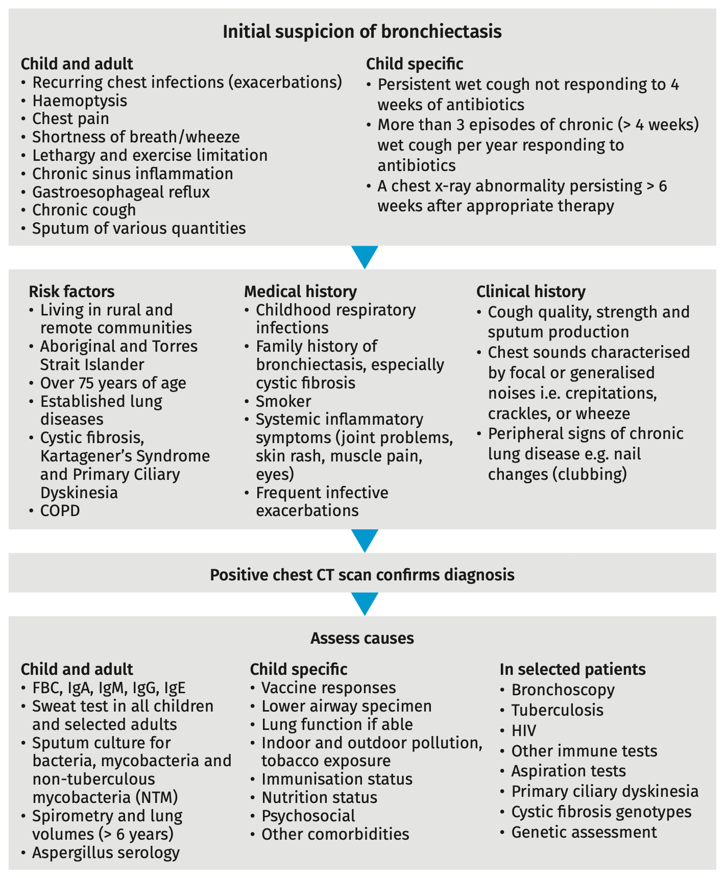
3. Management of bronchiectasis 1
- Management involves improving mucus clearance, while reducing airway bacterial colonisation, inflammation, and structural damage by:
- minimising symptoms (i.e. cough)
- reducing hospital admissions
- preventing lung infections
- improving quality of life
- improving exercise tolerance
- maintaining lung function
- reducing frequency and severity of exacerbations
- prolonging survival
- aggressively identifying and managing comorbidities, including:
- Chronic obstructive pulmonary disease
- severe respiratory infections
- GORD
- Asthma (adults and children > 12)
- chronic bronchitis
- Support patient self-management
- Discuss bronchiectasis and:
- airway clearance manoeuvres. See Resource 2.
- how to control breathlessness and Anxiety disorders
- develop an action plan. See 3.10 Action plan
- medicine usage, effects and adherence
- provide supportive resources. See Resource 3.
- If the patient also has COPD refer them to SMoCC, a phone service that supports patients manage their condition. See Resource 4.
- Encourage the patient to identify barriers to adequate lifestyle modification and
medical adherence and create goals to overcome those barriers. See Engaging our patients
- Discuss bronchiectasis and:
- Social-emotional support
- See Social-emotional wellbeing
- Smoking cessation 1–4
- Patients who stop smoking reduce the likelihood of lung infections and bronchiectasis progression
- See Smoking cessation
- Prevent respiratory infections 1–4
- Respiratory illnesses contribute to bronchiectasis exacerbations and progression
- Provide Influenza, pneumococcal and COVID-19 vaccines as per the Australian Immunisation Handbook
- Avoid environmental pollutants 2
- Patients should avoid environmental pollutants (see Table 1.) which can exacerbate:
- coughing
- sputum volume, consistency and purulence
- shortness of breath
- exercise intolerance
- fatigue
- haemoptysis
- Patients should avoid environmental pollutants (see Table 1.) which can exacerbate:
Table 1. Environmental pollutants to avoid in bronchiectasis 2 |
|---|
|
- Airway clearance technique 1–4
- Main therapy to clear excess lung secretions to improve ventilation and reduce hospital presentations
- Technique:
- start with 5 deep abdominal breaths. Expand chest fully, starting with the
diaphragm and lower ribs. Avoid lifting or shrugging shoulders - do 30–60 seconds of relaxed breathing. Breathe from the diaphragm. The patient should feel their stomach rising and falling with each breath. Shoulders should be kept as relaxed as possible
- do another 5 deep abdominal breaths
- follow this with 30–60 seconds of relaxed breathing
- take a medium sized breath in and huff the air out a little more forcefully
- start with 3 cycles of gentle huffs. Finish with 2 cycles of more forceful huffs
- finish with a cough to clear any secretions left in the main airways
- repeat above cycle 2–3 times or until no more secretions can be removed
- start with 5 deep abdominal breaths. Expand chest fully, starting with the
- Refer to a physiotherapist if patient is unable to clear lung secretions
- See Resource 2. for further information
- Improve physical activity tolerance 1–4
- Enhances airways clearance
- Should include moderate to high intensity aerobic exercises, strength training and mobility exercises
- Refer to an exercise physiologist for pulmonary rehabilitation or exercise program
- See Physical activity and sleep
- Pulmonary rehabilitation program
- An important hospital avoidance strategy offered to all patients with:
- poor physical activity tolerance
- > 2 exacerbations per year
- If no local program available:
- advocate for a service
- refer to the Pulmonary Rehabilitation Toolkit. See Resource 5.
- contact the chronic condition coordinator or the Lung Foundation for
rehabilitation program details and training. See Resource 6.
- An important hospital avoidance strategy offered to all patients with:
- Nutrition
- Lung disease increases the risk of poor nutrition, weight loss and reduced muscle strength because of:
- increased energy needs
- decreased appetite
- lack of energy to shop, cook or eat meals
- an increased need for certain vitamins, minerals and antioxidants
- Refer to MO/NP or dietitian if patient has unintended weight loss or weight gain
- See Diet and nutrition
- Lung disease increases the risk of poor nutrition, weight loss and reduced muscle strength because of:
- Action plan 2
- Develop an action plan (Resource 7.) with the patient so they can:
- recognise and monitor exacerbations and severity. See Table 3.
- intervene early to prevent exacerbations
- understand and feel comfortable using it
- Review and update action plan each visit, especially when changing medicines. See Table 2.
- Develop an action plan (Resource 7.) with the patient so they can:
Table 2. Bronchiectasis action plan | |
|---|---|
When feeling well | |
|
|
If ≥ 3 of these symptoms:
| Action
|
When feeling very unwell
| Action
|
4. Medicines for bronchiectasis
- Sputum sample 1–4
- Always take sputum samples and treat early
- Bronchiectasis patients often have positive sputum culture results. This does not mandate antibiotic use unless:
- the patient has an exacerbation (see Table 3.) or
- results show a new isolation of P. aeruginosa
- Exclude NTM infection by collecting at least 3 sputum samples for mycobacterial culture, in all patients before azithromycin use
Table 3. Identifying an exacerbation and severity 1–3 | ||
|---|---|---|
Key symptoms | Severe | Very severe |
|
|
|
Severe bronchiectasis exacerbations are similar to pneumonia. Exclude with a chest x-ray | ||
- Eradication of Pseudomonas aeruginosa (P. aeruginosa) 1–4
- The presence of P. aeruginosa in the airways is associated with increased:
- exacerbations
- risk of hospitalisation
- risk of mortality
- If a patient is clinically stable when P. aeruginosa is first identified, do not treat. Consult the Antimicrobial Stewardship (AMS) or a respiratory specialist to avoid promoting antibiotic resistance
- The presence of P. aeruginosa in the airways is associated with increased:
- Long-term antibiotics to reduce exacerbation frequency and symptoms in adults 1,2,4
- Seek advice from AMS or a respiratory specialist if a patient has:
- ≥ 6 exacerbations over 12 months or
- ≥ 2 hospitalisations over 12 months or
- > 6 months of continuous symptoms
- Routine long-term (6–12 months) oral or nebulised antibiotics are not recommended as antibiotic resistance is a common outcome
- Seek advice from AMS or a respiratory specialist if a patient has:
- Azithromycin prophylaxis in children with non-cystic fibrosis (non-CF) bronchiectasis or chronic suppurative lung disease (CSLD) 6,7
- Prior to initiation of azithromycin as maintenance therapy, the following are required:
- child has been reviewed by a respiratory consultant
- presence of bronchiectasis or CSLD
- ≥ 3 exacerbations and/or ≥ 2 hospitalisations in previous 12 months
- failed trial of long-term non-macrolide antibiotics for at least three months
- documented evidence of NTM exclusion in the lower airways
- non-pharmacological interventions are optimised and adhered to
- documented baseline liver function test and ECG
- Azithromycin is not initiated if:
- evidence of NTM infection
- allergy to macrolides
- abnormal liver function test
- medicine interactions e.g. antiarrhythmics
- See Table 4. for dosing and follow-up in children
- Prior to initiation of azithromycin as maintenance therapy, the following are required:
Consult specialist and hospitalise any patient with severe exacerbations with chronic P. aeruginosa colonisation or those in MRSA prevalent communities
Table 4. Long-term azithromycin (non-LAM) dosing schedule in children 6,7 | |
|---|---|
< 25kg weight |
|
25–40kg weight |
|
> 40kg weight |
|
Follow-up | |
| |
Table 5. Other medicines for bronchiectasis 1–4 |
|---|
Smoking cessation medicines |
|
Oxygen therapy |
|
Table 6. Medicines to treat adults with bronchiectasis 2–4,6 |
|---|
For severe and non-severe exacerbations without chronic P. aeruginosa colonisation
|
|
For adults with severe exacerbations without chronic P. aeruginosa colonisation where above oral therapy is inadequate
|
|
For non-severe exacerbations with chronic P. aeruginosa colonisation
|
Table 7. Medicines to treat children > 1 month of age with bronchiectasis 7 | |
|---|---|
For first or new isolation of P. aeruginosa colonisation without exacerbation | |
|
|
|
|
For first or new isolation of P. aeruginosa colonisation with exacerbation | |
| |
For chronic P. aeruginosa colonisation with exacerbation | |
| |
|
|
For acute exacerbations without P. aeruginosa colonisation
| |
|
|
|
|
For recurrent exacerbations without P. aeruginosa colonisation | |
| |
5. Cycle of care
Cycle of care summary for bronchiectasis | ||
|---|---|---|
Action | Dx | Review frequency |
Height | - | |
Blood pressure | 2 yrly | |
Weight | 2 yrly | |
BMI | 2 yrly | |
Pulse rate | 2 yrly | |
Respiratory rate | 2 yrly | |
Temperature | 2 yrly | |
Spirometry > 6 years age | Minimum 12 mthly (adults) 6 mthly (children) | |
Oxygen saturations | Minimum 12 mthly (adults) 6 mthly (children) | |
FBC | 12 mthly | |
IgG, IgA, IgM, IgE | - | |
Sweat test | In all children and select adults | |
Sputum culture | Minimum 12 mthly (adults) 6 mthly (children) | |
Aspergillus serology | - | |
Lifestyle modifications education | Every visit | |
Social-emotional wellbeing | 12 mthly | |
Bronchiectasis action plan | 12 mthly | |
Influenza, pneumococcal and COVID-19 vaccines | Recommended. See the Australian Immunisation Handbook for schedule | |
Chest x-ray | During chest infection to rule out pneumonia | |
High resolution CT | - | |
Medicine review | Each visit | |
Self monitoring (action plan) | Minimum 12 mthly (adults) 6 mthly (children) | |
HW/RN review | Ongoing monitoring with recall register | |
MO/NP review | Minimum 12 mthly (adults) 6 mthly (children) | |
Pulmonary rehabilitation | PRN for poor physical activity tolerance | |
Physiotherapist | PRN for airway clearance manoeuvers and education | |
Specialist MO | 12 mthly (adults) 6 mthly (children) | |
6. References
- All Chronic Conditions Manual references are available via the downloadable References PDF
7. Resources
- Bronchiectasis prediction tools for predicting mortality and exacerbation rates in non-cf bronchiectasis
- Airway clearance manoeuvres resources
- Bronchiectasis patient resources
- Self-Management of Chronic Conditions (SMoCC) service
- The Australian Lung Foundation Pulmonary Rehabilitation Toolkit
- The Lung Foundation training and education website
- A bronchiectasis action plan